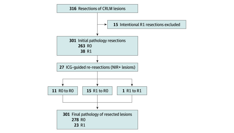
Figure 2. Flow Diagram of Changes in Final Pathology After Re-Resection.
Per-lesion analysis of all resected and histologically proven colorectal liver metastasis (CRLM) lesions showing an initial radical resection rate of 82.3% and final radical resection rate of 92.4% after additional resection. Based on fluorescence signal with histopathologic assessment as the gold standard, the accuracy of indocyanine green (ICG)–fluorescence imaging as an indicator for resection margins was calculated in the final step. NIR+ indicates positive near-infrared fluorescence signal; R0, negative tumor margin; R1, positive tumor margin.

