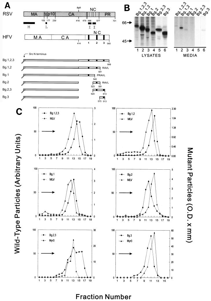
FIG. 4.
The HFV Gag protein contains at least two I domains. (A) RSV and HFV Gag proteins are illustrated. The vertical dotted lines separate HFV Gag into regions which are analogous to the cleavage products of other retroviral Gag proteins. The black vertical bars in HFV NC indicate the position of the three GR boxes. The numbers below the chimeric proteins indicate the HFV Gag residues contained in the constructs and the letters at the end of the molecules are the single letter codes of any nonviral residues present. (B) Expression of RSV-HFV chimeras was performed as described in the legend for Fig. 1 except that l-[35S]methionine was used to label the cells, and the relevant proteins were immunoprecipitated with α-RSV antibody. (C) Sucrose gradient analysis was performed as described in the legend for Fig. 1 except that the RSV-HFV chimeras were labeled with l-[35S]methionine and immunoprecipitated with α-RSV antibody, and two constructs (Bg.2,3 and Bg.3) were mixed with labeled RSV Gag-only particles (rather than authentic MLV) before layering on the gradient.

