FIG. 1.
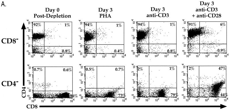
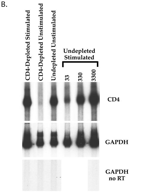
(A) Stimulation of CD8-depleted (top row) and CD4-depleted (bottom row) PBL. PBL were depleted by panning and stimulated with either PHA, anti-CD3 MAb, or anti-CD3 and anti-CD28 MAbs. Cells were analyzed by flow cytometry for expression of CD4 (red 613) and CD8 (allophycocyanin) following 3 days of stimulation. CD4+/CD8+ cells were not observed when cells were stimulated with anti-CD28 alone (data not shown). The percentage of cells in each quadrant is shown. (B) CD4 mRNA expression in costimulated and unstimulated CD4-depleted leukocytes. PBL were depleted of CD4+ cells by panning and stimulated with anti-CD3 and anti-CD28 MAbs or cultured unstimulated in parallel. Three days later, RNA was purified from the CD4-depleted and undepleted populations and subjected to RT-PCR for CD4 (top panel) and GAPDH (middle panel). Standards consisting of dilutions of undepleted stimulated PBL were amplified in parallel and are indicated by number of input cell equivalents. The primers for CD4 mRNA amplify a region containing an RNA splice site and therefore do not amplify potentially contaminating chromosomal DNA sequences. A “no RT” control was performed for GAPDH (bottom panel) to detect the presence of contaminating DNA sequences, of which there were none. The ratio of the level of CD4 mRNA signal to GAPDH signal was 22-fold higher in the costimulated CD8+ population than in the unstimulated CD8+ population.


