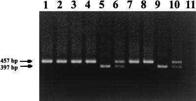
FIG. 4.
PCR detection of viral sequences from lymph node biopsies 19 weeks after challenge of vaccine group 1 with SHIVKU-1. Lymph node biopsies were performed; DNA was extracted and used in nested PCR with oligonucleotides that amplified the vpu gene of SHIVKU-1 as described in the text. Aliquots of the nested PCR were run on a 1.5% agarose gel and stained with ethidium bromide. Lanes 1 to 7, amplification of vpu sequences from lymph node DNA of macaques 42105, 42107, PLk, PPm, PDj, PNa, and PFy, respectively; lane 8, amplification of vpu sequences from plasmid p3′SHIV (positive control for full length vpu gene); lane 9, amplification of vpu sequences from plasmid pDSDvpu (positive control for truncated vpu gene); lane 10, amplification of vpu sequences from plasmids p3′SHIV and pDSDvpu; lane 11, amplification of vpu from uninfected PBMC DNA (negative control).

