
Fig. 1.
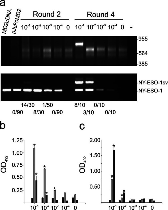
a Biopanning with anti-NY-ESO-1 antibodies diluted in IgG from healthy human individuals. Top panel: ethidium bromide stained agarose gel of EcoRI/XhoI digests of pJuFoMD2 library (lane 2 from left), selected phagemids after round 2 (lanes 3–7) and round 4 (lanes 8–12). Empty lanes in 1 and 13. Lower panel: PCR for NY-ESO-1 cDNA in MD2 tumor cDNA (10 ng, positive control, lane 1), pJuFoMD2 library (50 ng, lane 2), selected phagemids (50 ng) after round 2 (lanes 3–7) and round 4 (lanes 8–12). PCR without template (lane 13). Figures above top panel denote molar fraction of NY-ESO-1-specific antibodies, figures below lower panel denote fraction of NY-ESO-1-encoding clones. Size markers are given to the right. b ELISA with sera in a and phages displaying NY-ESO-1 (dark gray) or NY-ESO-1sv (light gray) and empty pJuFo phages (black). Vertical lines denote standard error for 4 experiments. Reactivity significantly different from empty pJuFo phages and normal human IgG are highlighted by * (p<0.05, Student’s t-test). c ELISA with sera in a and recombinant NY-ESO-1 (dark gray) or NY-ESO-1sv (light gray) proteins and negative control protein p352 (black)