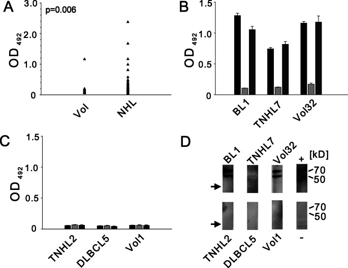
Fig. 1A–D.
Analysis of anti-ATF-2 antibodies in sera from healthy volunteers and patients with non-Hodgkin’s lymphoma (NHL). A Results of enzyme-linked immunosorbent assay (ELISA) using recombinant ATF-2 as target antigen. Arrow depicts mean of duplicate experiments for each serum from healthy volunteers (Vol) and patients (NHL). OD 492, optical density at 492 nm. B, C Results of competition ELISA shown for individual positive (B) and negative (C) sera. Bars denote results in ELISA (black), after preincubation of serum with recombinant ATF-2 (light grey) or irrelevant protein p352 (dark grey) with error bars representing standard error of the mean of duplicate experiments. BL Burkitt’s lymphoma, T-NHL T-cell lymphoma, DLBCL diffuse large B-cell lymphoma. D Western blot analysis of individual sera from B and C. Expected protein bands corresponding to recombinant ATF-2 are indicated at 70 and 50 kDa. No reactivity to p352 (arrow) is seen. Lane titled “+” represents experiment using anti-ATF-2 primary antibody and appropriate secondary antibody (see text). Lane titled “−” shows reactivity of BL1 serum toward a lysate of E. coli M15 cells prepared in parallel with lane BL1

