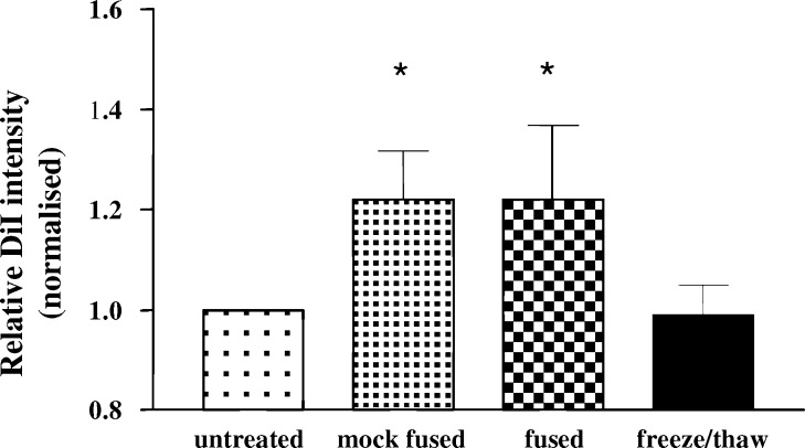
Fig. 4.
Uptake of tumour cells by DC. Dye labelled SW620 tumour cells (DiI) were either untreated, mock fused, fused or freeze/thawed prior to co-culture (18 h) with DC. Following co-culture, DC were labelled with HLA-DR and the MFI of the DC associated DiI fluorescence was determined by FACS analysis. Results are expressed as the change normalised relative to DC co-cultured with untreated tumour cells (1.0). Data are from five separate experiments and are shown as mean±SEM. * indicates P<0.05 following comparison with untreated cells

