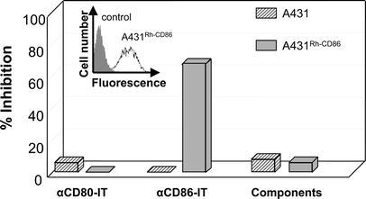
Fig. 2.

Cytotoxicity of αCD86-IT and αCD80-IT to RhCD86-transfected A431 cells. A431 cells transfected with RhCD86 were stained with anti-CD86 (clone 1G10) or control IgG followed by rabbit antihuman FITC-conjugated IgG. Results were analyzed by flow cytometry as indicated in the inserted figure. Both RhCD86 and mock transfected A431 cells were incubated with 10-8M αCD86-IT, αCD80-IT or unconjugated MAbs plus free gelonin for 72 h and additional 16 h with 3H-leucine. Inhibition of protein synthesis is expressed as percentage of 3H-leucine incorporation by untreated cells. Concentrations refer to the amount of gelonin in the IT used
