
Fig. 6.
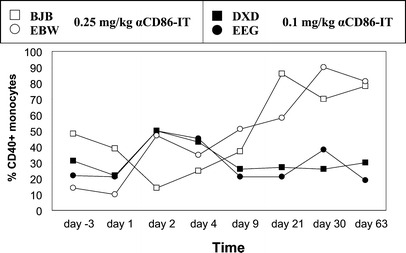
Expression of CD40 on monocytes of αCD86-IT–treated rhesus monkeys. PBMCs from rhesus monkeys were stained with anti-CD14 conjugated with biotin and anti-CD40 conjugated with PE, followed by streptavidin-FITC. Depicted is the percentage of CD40+ cells with in CD14 gate, as analyzed by flow cytometry