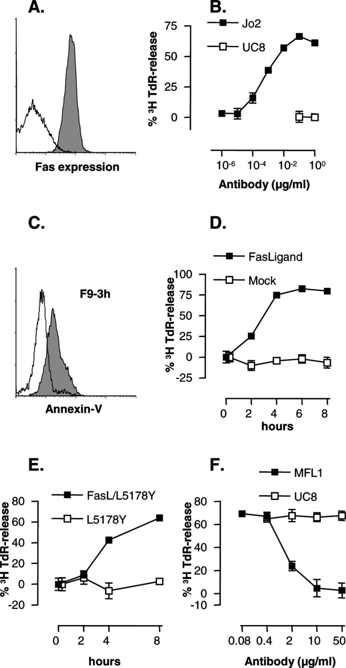
Fig. 1a–f.

Id+ F9 cells express Fas and rapidly undergo apoptosis when exposed to a variety of ligands that bind Fas. a Staining of F9 cells with anti-Fas mAb Jo2 (shaded histogram) and control mAb UC8 (open histogram). b Anti-Fas mAbs induce apoptosis of [3H]TdR-labeled F9 cells and release of labeled nucleosomal fragments of DNA in a 6-h assay. c Annexin V staining of F9 cells exposed to FasL-CD8 fusion protein (shaded) or SN from mock-transfected COS cells in a 3-h assay (open). d Kinetics of apoptosis of F9 cells induced by FasL-CD8 fusion protein in a [3H]TdR-release assay. e Kinetics of apoptosis of F9 induced by FasL-transfected L5178Y (E/T=10:1) in a [3H]TdR-release assay. f Anti-FasL mAb MFL1 inhibits killing of [3H]TdR-labeled F9 target cells exposed to mouse FasL-transfected L5178Y (E/T=10:1) in a 6-h assay
