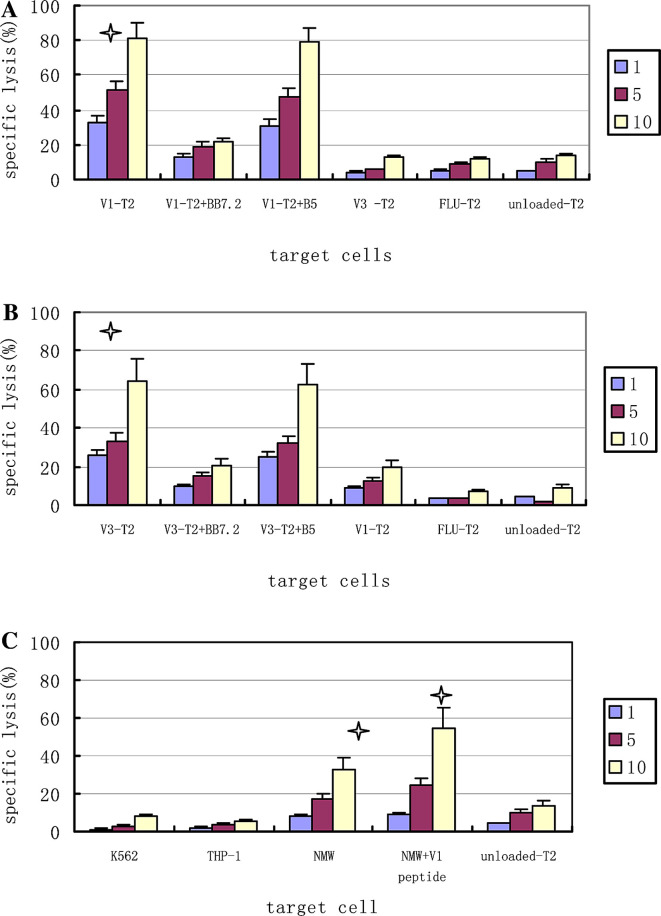
Fig. 4.
CTL cytotoxicity Cytotoxicity was determined in a standard LDH assay at E/T ratios of 1:1 and 5:1 and 10:1. a Effector T cells induced by IgHV1 (QLVQSGAEV) peptide stimulations, against T2 cells loaded with IgHV1, IgHV3, FLU peptide as target cells, preincubation with anti-HLA-A2 mAb BB7.2 and the isotype mAb B5. b Effector T cells induced by IgHV3 (SLYLQMNSL) peptide stimulations, against T2 cells loaded with IgHV1, IgHV3 or FLU peptide as targets cells and preincubation with BB7.2 and the isotype mAb B5. c Effector T cells induced by IgHV1 (QLVQSGAEV) peptide stimulations, against some tumor cell lines: NAMALWA (HLA-A*0201-positive, IgHV1-positive), THP-1 (HLA-A*0201-positive, IgHV1-negative, K562 (HLA-A*0201-nagative, IgHV1-negative), Data are expressed as the mean ± M of three independent experiments, One-way ANOVA test was used to test the significance of the specific lysis of the CTLs, and the lysis of the peptides-unloaded T2 cells as the target cells was used as the control group to compare with the lysis of the different peptides-loaded T2 cells groups as the target cells.*Significant lysis (P<0.05). V1: IgHV1 (QLVQSGAEV) peptide; V3: IgHV3 (SLYLQMNSL); FLU: irrelevant influenza matrix-derived peptide (GILGFVFTL); NAM: NAMALWA cell

