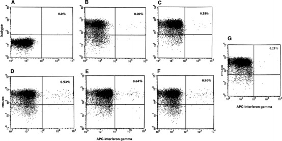
Fig. 3A–G.

Intracellular cytokine analysis. CD8+ cells from PBL were isolated by positive selection using Dynal magnetic beads. Cells were stimulated with PSA-peptide-pulsed autologous DC for 6 h. Cells were stained with anti-CD8 antibody conjugated with FITC (Y axis) and then stained for intracellular staining with antihuman IFN-γ conjugated with APC (X axis) according to the method described. A Isotype matched control. B Cells stimulated with DC only. C Cells stimulated with PSA-1 peptide-pulsed DC. D Cells stimulated with PSA-2 pulsed DC. E Cells stimulated with PSA-3-pulsed DC. F Cells stimulated with PSA-4-pulsed DC. G Cells stimulated with Mart-1 peptide-pulsed DC
