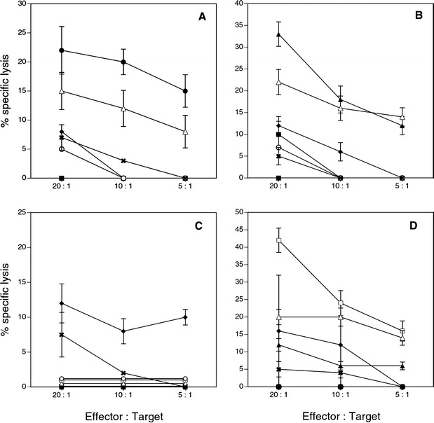
Fig. 5A–D.

Cytotoxicity assay with in vitro expanded peptide-specific CTL. CD8+ cells were stimulated in vitro with matured autologous DC pulsed with one of the PSA-derived peptides. Cytotoxicity was measured by 51Cr release from labeled different target cells. Target cells were T2 cells alone, T2 cells pulsed with one of the four PSA peptides, T2 cells pulsed with Flu peptide, HLA-A2 positive and PSA positive prostate cancer cell line LN CaP, and HLA-A2 positive and PSA negative melanoma cell line CS-M. A CTL activity with responding CD8+ cells stimulated with DC pulsed with PSA-1. B CTL activity with responding CD8+ cells stimulated with DC pulsed with PSA-2. C CTL activity with responding CD8+ cells stimulated with DC pulsed with PSA-3. D CTL activity with responding CD8+ cells stimulated with DC pulsed with PSA-4. Solid square % specific lysis of the target T2 cells alone. Solid circle % specific lysis of the target T2 cells pulsed with peptide PSA-1. Solid triangle % specific lysis of the target T2 cells pulsed with peptide PSA-2. Solid diamond % specific of the target T2 cells pulsed with peptide PSA-3. Open square % specific lysis of the target T2cells pulsed with peptide PSA-4. Open circle % specific lysis of the target T2 cells pulsed with Flu peptide. Open triangle % specific lysis of the target LN CaP tumor cells. X % specific lysis of HLA A2 positive melanoma tumor target CS-M cells
