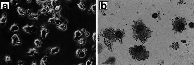
Fig. 1a, b.

Morphological features of DCs derived from PLC patients. Cultured DCs were monitored by light microscopy before administration (a). DCs from PLC patients morphologically appeared to be immature with short, blunt prolongations, despite stimulation with TNF-α(b) (May-Giemsa staining, ×200)
