
Fig. 2A, B.
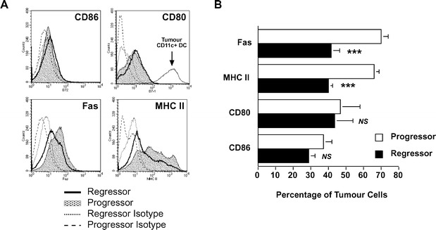
Progressor tumour cells express higher levels of MHC class II and Fas but negligible amounts of CD80 or CD86 costimulatory molecules. A Two-colour flow cytometry was used to identify tumour cells as CD45− along with a second marker against CD86, CD80, Fas or MHC II. Both progressor (shaded histogram) and regressor (thick-lined histogram) tumour expression of the markers are shown. Isotype-matched control antibodies for both progressor (dashed line) and regressor (dotted line) are indicated. The fluorescence intensity of CD80 on CD11c+CD45+ regressor infiltrating dendritic cells (as an example of APC expression of CD80) is shown by the thin solid line in the corresponding histogram. B Statistical analysis by a paired Student's t-test revealed that a significantly greater percentage of progressor tumour cells (open bars) expressed MHC II and Fas than regressor tumours (solid bars) (***P<0.001, n=7). There was no significant difference with respect to CD80 or CD86 expression