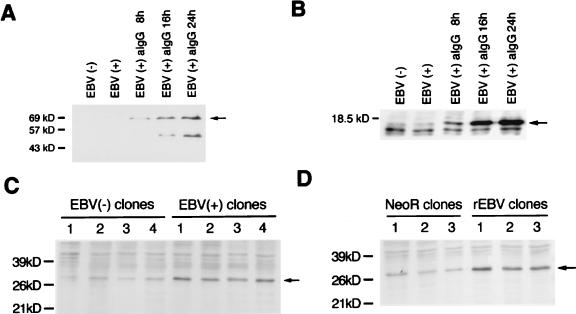
FIG. 5.
Western blot analysis of LMP1, BHRF1, and bcl-2 protein. (A and B) LMP1 (A) and BHRF1 (B) (arrows) are detected only after cells are treated with anti-IgG, which induces the virus lytic cycle, indicating that these proteins are lytic gene products rather than latent gene products. Minor bands appear to be degraded proteins. (C) bcl-2 protein expression (arrow) of EBV-positive and -negative cell clones derived from parental Akata cells (four clones each). (D) bcl-2 protein expression (arrow) of EBV-reinfected and neoR-transfected clones derived from an EBV-negative Akata clone (three clones each). bcl-2 protein is expressed at a higher level in EBV-infected clones than in uninfected clones. Molecular masses of protein markers are shown at the left of each panel.

