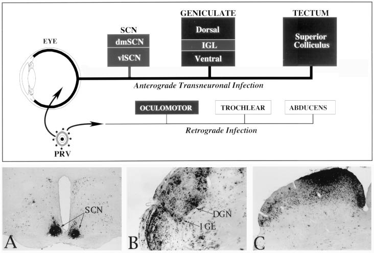
FIG. 1.
A schematic illustration of the organization of the neuronal circuitry that was the subject of analysis in this investigation is shown at the top of the figure. Filled boxes represent areas that were included in the analysis, while open boxes represent motor nuclei that were virus infected but not subjected to analysis in this study. Injections of virus into the vitreous body of the eye produced an anterograde transynaptic infection of retinorecipient neurons in the diencephalon (SCN and geniculate complex) and midbrain (tectum). The distribution of infected neurons in these regions at long postinoculation intervals after injection of PRV Becker is illustrated in the photomicrographs of the SCN (A), the geniculate complex that includes the DGN and IGL (B), and the tectum (C). Leakage of virus into the eye also produced a retrograde infection of motor neurons innervating eye musculature (oculomotor, trochlear, and abducens nuclei). dmSCN, dorsomedial suprachiasmic nucleus; vlSCN, ventrolateral suprachiasmic nucleus.

