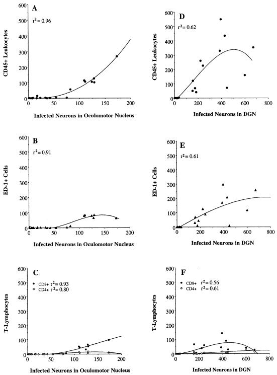
FIG. 6.
Quantitative analyses showing the relation between leukocyte infiltration and viral infection in the oculomotor nucleus and DGN after injection of PRV Becker into the vitreous body of the eye. CD45-, ED-1-, CD8β-, and CD4-immunopositive leukocytes and neurons containing immunoreactivity to viral structural proteins were counted in brain tissue from each virus-infected rat. The individual data points in these figures are the actual number of cells counted for each rat in each region of infection. These figures also show the best-fitting curvilinear line of the data. No inferences should be made about the shape of the end of each curve, which is based on only a few data points. r2 indicates the strength of the association between leukocyte infiltration and viral infection. Note that the range of values on the x axis changes according to the area of infection.

