
Fig. 2.
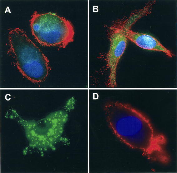
Uptake of irradiated A549 and primary lung tumor cells by DC. Immature DC were loaded with PKH67-labeled, irradiated A549 or primary lung tumor cells for 24 h and then transferred to glass coverslips. Adherent DC were stained with Alexa Fluor 568-conjugated anti-CD11c (red) and the nuclear stain DAPI (blue). Samples were analyzed with a widefield epifluorescent/brightfield microscope. A A549 cell material internalized by DC; B primary lung tumor cell material internalized by DC; C A549 cell alone; D DC alone. (mid-plane images)