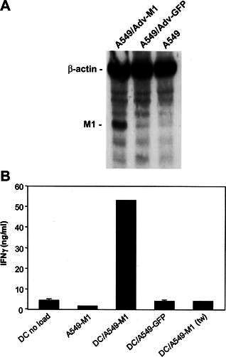
Fig. 5.

DC loaded with irradiated A549-M1 tumor cells stimulate IFNγ release by antigen-specific T cells. A Western blot analysis of M1 protein expression by A549-M1 cells. Lysates from equivalent number of control A549 cells and A549 cells infected with an adenovirus expressing influenza M1 protein (Adv-M1) or green fluorescent protein (Adv-GFP) were electrophoresed on a 12% SDS-PAGE, blotted onto an Immobilon-P PVDF membrane and hybridized with anti-influenza-matrix-HRP and anti-human β-actin antibodies simultaneously. The M1 and β-actin proteins on the membrane were visualized with the LumiGLO Chemiluminescent Substrate followed by exposure of the membrane to Kodak BioMax Light film. B Immature DC were loaded with or without an equal number of A549-M1 or A549-GFP tumor cells for 24 h. The cells were then matured with BCG and IFNγ and cultured with an equal number of cells from a M1-peptide-specific T cell line. After 24 h, supernatants were collected and analyzed for IFNγ release by ELISA. Cultures in which DC were separated from tumor cells by a 0.4-mm transwell membrane during the loading period are indicated by the notation ( tw). Bars represent the mean ± SD of triplicate wells. The results are representatives of four ( A) and three ( B) experiments
