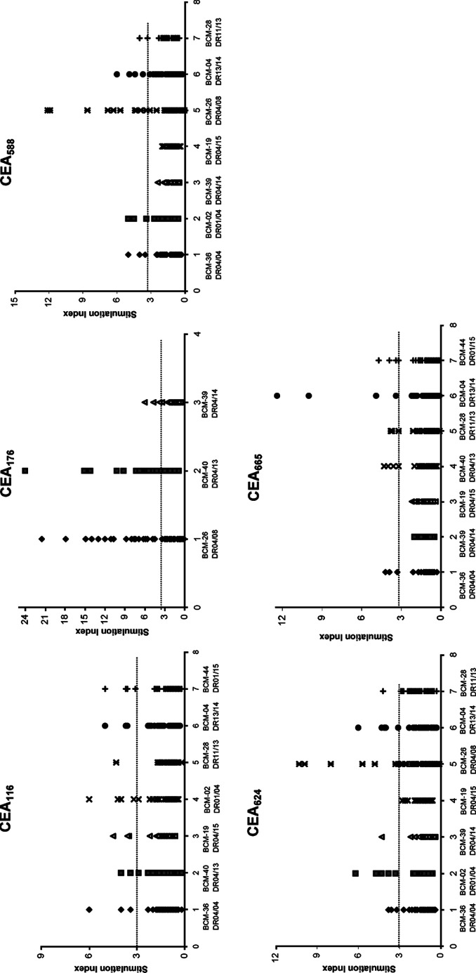
Fig. 1.
Initial screening of T-cell responses to predicted peptides from carcinoembryonic antigen (CEA). Peripheral blood mononuclear cells (PBMCs) from healthy donors with HLA-DR4 and other HLA-DR genotypes were seeded on 96-well plates and stimulated with each peptide (20 μg/ml) for 1 week. [3H]-thymidine incorporation of the primed T-cells (2×10⁵ cells/well) was measured after restimulation with autologous PBMCs (1×10⁵ cells/well) as antigen-presenting cells with or without the corresponding peptides (20 μg/ml). Responses to each peptide were tested in total 48-wells. Wells were scored positive if the counts per minute of T-cells stimulated with peptides was >1000 and stimulation indices (SI) >3 [31, 40, 48, 64]. The results are reported as SI of each tested well of different donors

