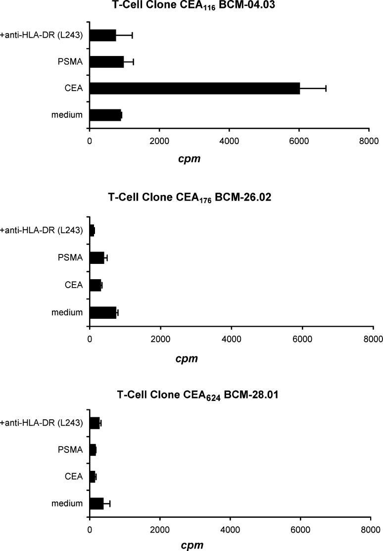
Fig. 6.
The CD4+ T-cell responses to natively processed CEA. Each of three peptide-reactive T-cell clones (CEA 116, CEA 176, and CEA 624; 3×10⁴/well) were stimulated in triplicate wells with irradiated autologous PBMC-derived DC (1.5×10³/well) pulsed with recombinant CEA protein (10 μg/ml) in the presence or absence of the anti-HLA-DR antibody (L243; 20 μg/ml). These T-cell clones were also stimulated with DC pulsed with irrelevant recombinant prostate-specific membrane antigen (PSMA) protein (10 μg/ml). T-cell proliferation was determined by [3H]-thymidine incorporation assay during the last 16 of 72 h of culture. The proliferation of T-cells coculture with CEA-pulsed DC was significantly higher than proliferation of T-cells with non-pulsed DC (medium control) and with addition of anti-HLA DR antibody (p<0.01). Values shown are the means of triplicate determinations (bars: SD). The representative result of one of three repeated experiments is shown

