
Fig. 3.
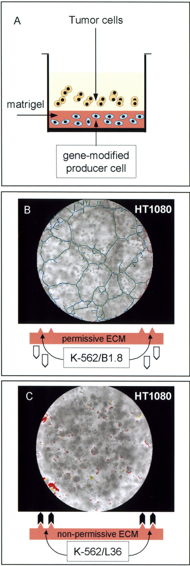
A Scheme of the modified Matrigel tube formation assay. The bottom of each plate well was coated with a diluted Matrigel (7 mg/ml) solution containing 5×10³ gene-modified K-562 cells (K-562/B1.8 or K-562/L36) and then allowed to solidify at 37°C for 30 min. After 96 h, between 103 and 5×10³ tumor cells were plated onto the gellified matrix and incubated for an additional 16-h period. B and C Light microscope photos of the human fibrosarcoma cell line HT-1080, plated for 16 h on either a B1.8-rich (B) or L36-rich (C) Matrigel substratum. Images were processed using software designed to detect tube formation [25]. One of three similar experiments is shown