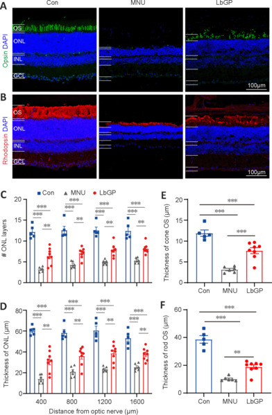
Figure 4.

LbGP improves photoreceptor structure and survival in the MNU-injured retina.
(A, B) Representative images of retinal sections from the intermediate region of each group. Opsin (Alexa FluorTM 488, green; A) and rhodopsin (Alexa FluorTM 647, red; B) immunostaining was used to label the outer segment (OS) of the cones and rods, respectively. Areas of retinal opsin and rhodopsin staining formed elongated or columnar shapes in the normal control (Con) group, dots or clumps in the MNU group, and long stripe shapes in the LbGP group retina. The nuclei were stained with 4,6-diamino-2-phenyl indole (DAPI; blue). Scale bars: 100 μm. (C, D) Quantification of the cell layers (C) and thickness (D) of the outer nuclear layer (ONL). In each group, retinal sections were evaluated from the center to the peripheral regions. (E, F) Thickness of the OS of cones (E) and rods (F) in each group. The ONL and OS measurements of the cones and rods were significantly thinner in the retinas of the MNU group than those in the Con group. Treatment with LbGP significantly increased these parameters. Data expressed as mean ± standard error of the mean (Con, n = 5; MNU, n = 6; LbGP, n = 8); **P < 0.01, ***P < 0.001 (one-way analysis of variance followed by Tukey’s post hoc test). Con: C57BL/6J mice; DAPI: 4′,6-diamidino-2-phenylindole; GCL: ganglion cell layer; INL: inner nuclei layer; LbGP: N-methyl-N-nitrosourea-injured mice with Lycium barbarum glycopeptide treatment; MNU: N-methyl-N-nitrosourea-injured mice without treatment; ONL: outer nuclei layer.
