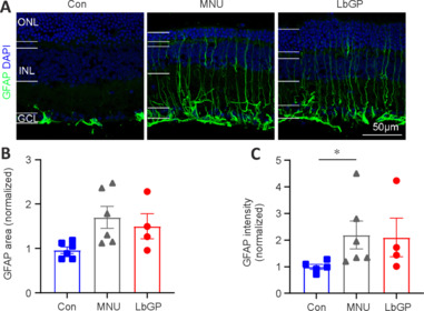
Figure 6.

LbGP hardly inhibits Müller glial cell activation in the MNU-injured retina.
(A) Representative images of retinal sections stained with GFAP (Alexa Fluor-488, green) and DAPI (blue) for each group. In both the untreated and LbGP-treated MNU-injured mice, GFAP expression in Müller glial cells was present throughout the retina. Scale bars: 50 μm. (B, C) Fluorescence area (B) and intensity (C) of GFAP-positive regions (normalized to the control) of each group. Data expressed as mean ± standard error of the mean (Con, n = 5; MNU, n = 6; LbGP, n = 4); *P < 0.05 (one-way analysis of variance followed by Tukey’s post hoc test). DAPI: 4′,6-Diamidino-2-phenylindole; GCL: ganglion cell layer; GFAP: glial fibrillary acidic protein; INL: inner nuclei layer; LbGP: N-methyl-N-nitrosourea-injured mice with Lycium barbarum glycopeptide treatment; MNU: N-methyl-N-nitrosourea-injured mice without treatment; ONL: outer nuclei layer.
