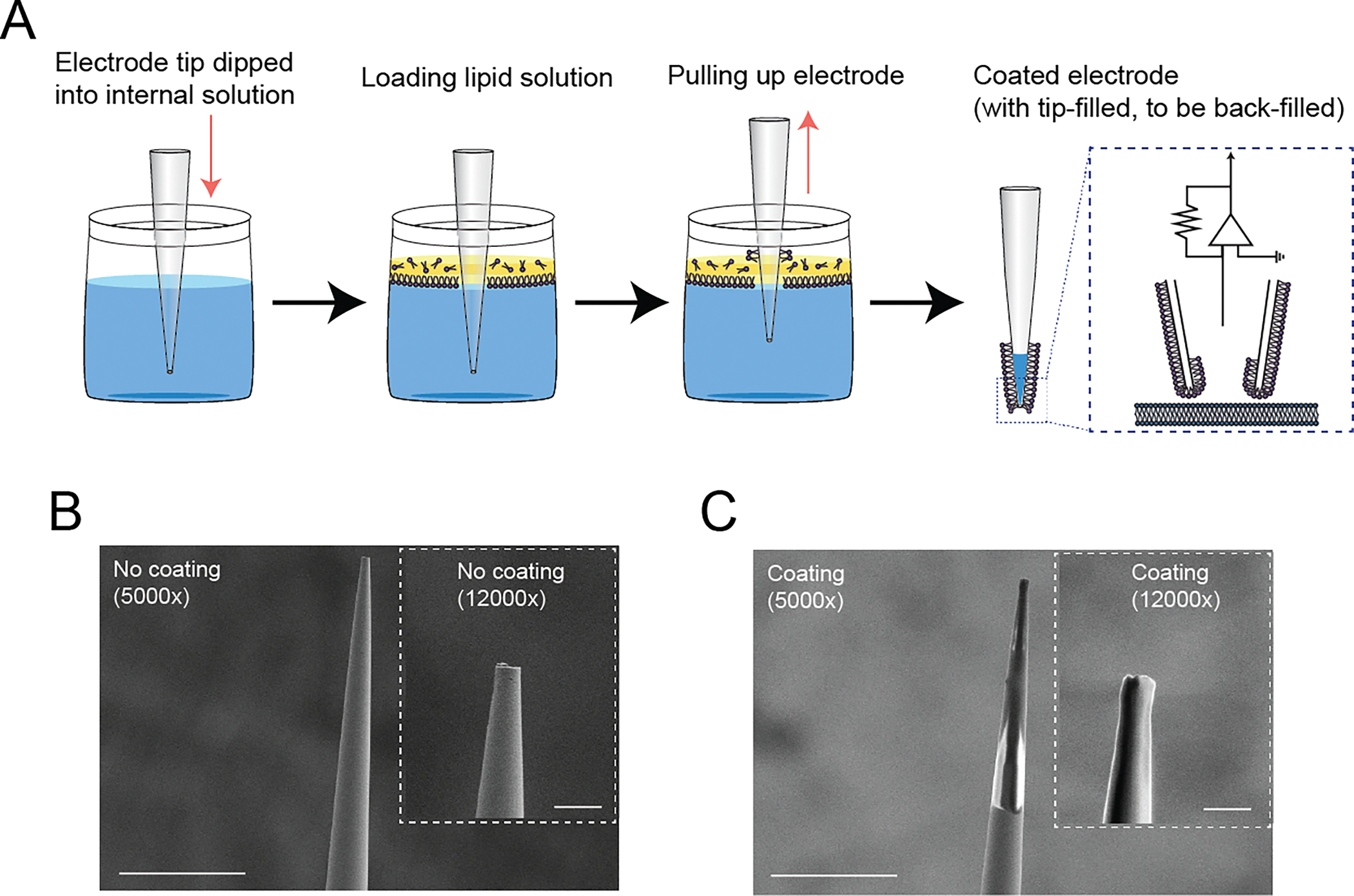
Figure 1.

Development of a membrane-coated glass electrode. (A) Graphical schematics of the coating process of the membrane-coated electrodes. The process is initiated by immersing the electrode tip into a reservoir containing a standard internal electrode solution. At the interface between the water and oil phases, a single phospholipid monolayer is expected to form (Noireaux and Libchaber, 2004). After adding a minimized layer of the prepared lipid solution onto the surface of the internal electrode solution, the electrode is vertically lifted using the micromanipulator. The resulting membrane-coated electrodes were promptly employed for electrophysiological recording, after the standard backfilling procedure with the internal electrode solution. (B) Surface visualization of an uncoated glass electrode at 5,000x magnification and 12,000x magnification (inset). Scale bar indicates 20 μm at 5,000x magnification and 2.5 μm at 12,000x magnification (inset). (C) Surface visualization of a membrane-coated glass electrode at 5,000x magnification and 12,000x magnification (inset). Scale bar indicates 20 μm at 5,000x magnification and 2.5 μm at 12,000x magnification (inset).
