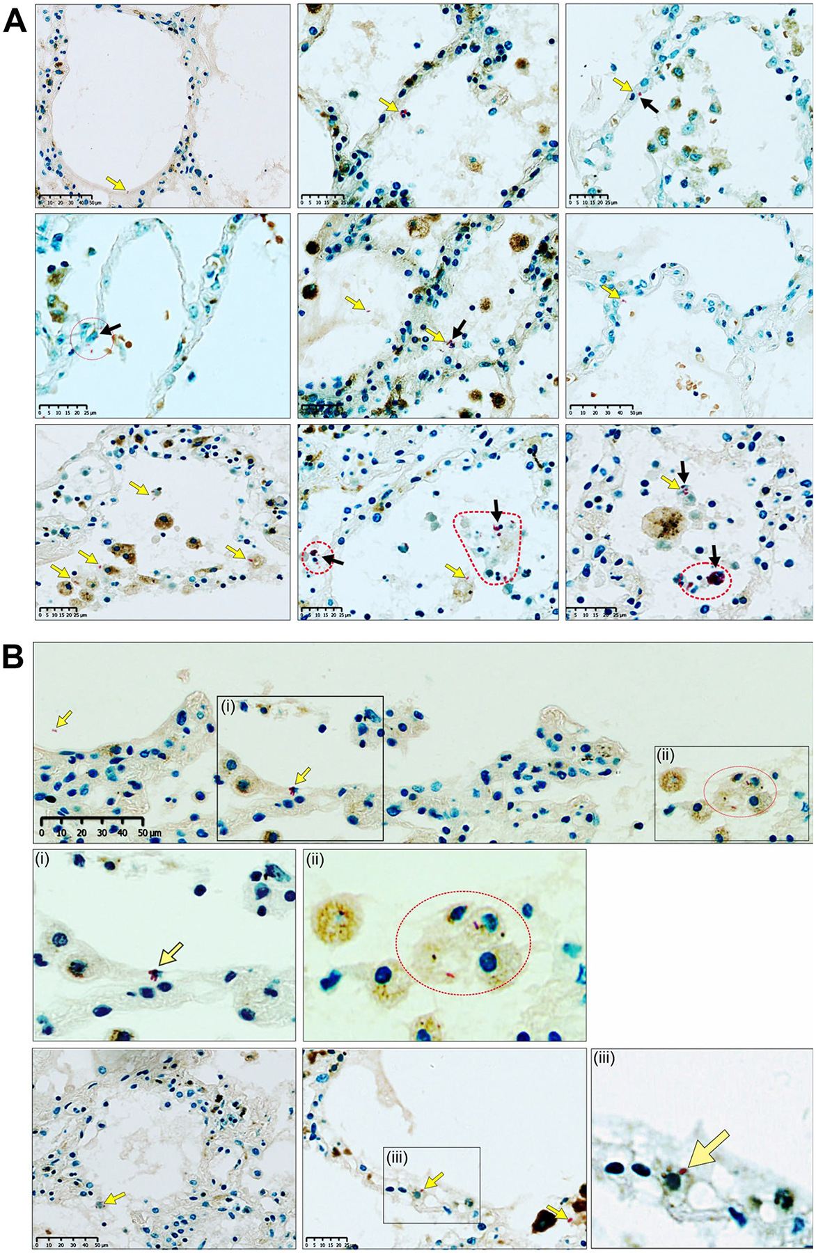
Fig. 10.

Visualization of M.tb within the alveoli and alveolar epithelium in the human lung. (A) Combined Ziehl-Neelsen(ZN)/CD68 and (B) ZN/Heme Oxygenase-1 (HO-1) staining of multiple sections of the lung parenchyma from a patient with microbiologically confirmed TB: Low-power images and high-power magnifications demonstrate acid-fast bacilli (AFBs) (yellow arrows) in extracellular and intracellular locations, including AFBs within alveolar epithelial cells (black arrows). Dotted red line shapes indicate M.tb-infected cells with multiple AFBs. The ZN method stains the AFBs of M.tb red in color, including cross-sections thereof that appear as red dot-like structures. CD68 is a macrophage immunomarker. A dilute HO-1 immunohistochemical stain permitted an improved visualization of cellular outlines. In (B), (i), (ii), and (iii) are magnifications. AFB = acid-fast bacilli; HO-1 = Heme-oxygenase 1; M.tb = Mycobacterium tuberculosis; TB = tuberculosis; ZN = Ziehl-Neelsen.
