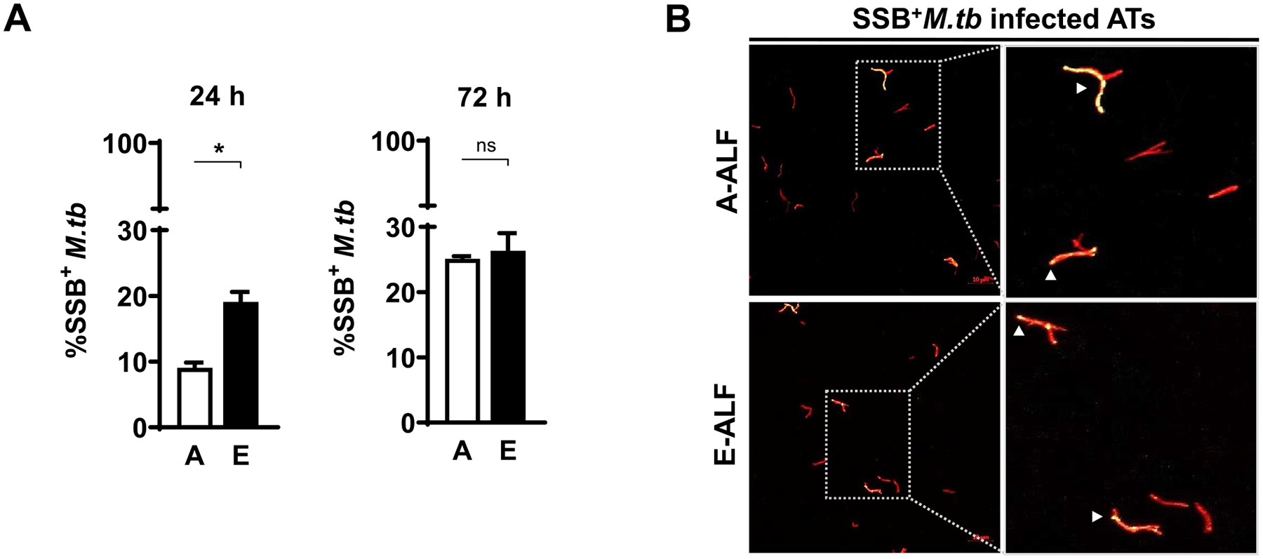
Fig. 2.

Elderly ALF-exposed M.tb has enhanced early replication during ATs infection. ATs were infected with the reporter SSB-GFP, smyc’:: mCherry M.tb strain at MOI of 10:1 and bacterial replication rate was determined by confocal microscopy at the indicated time points. (A) Percentage of SSB+M.tb exposed to A- and E-ALFs at 24 hpi and 72 hpi, n = 2–4 (mean ± SEM) in replicates, using two different A-ALFs and four different E-ALFs. (B) Representative confocal images of ATs infected with A- and E-ALF-M.tb at 72 hpi. The region indicated by a gray dashed line is shown expanded on the right (top panels, A-ALF; bottom panels, E-ALF). Replicating SSB+M.tb is indicated by white arrowheads, showing merged (yellow) foci. Events were enumerated by counting at least 50 independent events (≥ 50 bacteria). <scale bars represent 10 μm>. Student’s unpaired t test analysis of Adult versus Elderly, *p < 0.05. The “n” values represent the number of biological replicas using an individual ALF sample from different adult or elderly human donors. A = adult ALF-exposed M.tb; ALF = alveolar lining fluid; ATs = alveolar epithelial type cells; E = elderly ALF-exposed M.tb; GFP = green fluorescent protein; hpi = hours post-infection; MOI = multiplicity of infection; M.tb = Mycobacterium tuberculosis; ns: no significant differences; SEM = standard error of the mean; SSB = single-stranded DNA binding protein.
