Fig. 5.

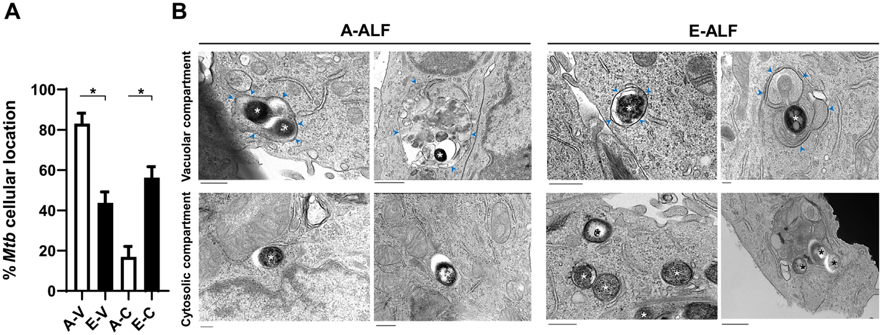
ALF-exposed M.tb drives differences in intracellular localization within ATs. ATs were infected with either A-ALF or E-ALF-exposed M.tb for 2 hours at MOI of 100:1 followed by 1 hour of gentamicin to kill extracellular M.tb. Relative proportion of intracellular bacteria located within membrane-bound vesicles or free in the cytosol by TEM. (A) Coded samples were scored by blinded analysis to quantify A-ALF-exposed and E-ALF-exposed M.tb located in vacuolar (endosomal/lysosomal) or cytosolic compartments at 72 hpi, n = 2 (mean ± SEM). (B) TEM micrographs of ALF-exposed M.tb. A-ALF-exposed and E-ALF-exposed M.tb were scored as “cytosolic, C” if they were not enclosed within a membrane or scored as “vacuolar, V” if they were surrounded by a vacuolar membrane. Vacuole membranes are indicated by blue arrowheads. Bacteria are indicated by asterisks. Values were determined by counting at least 100 independent events (bacteria). Student’s unpaired t test analyses of A-V versus E-V and A-C versus E-C; *p < 0.05. A-V: adult ALF-exposed M.tb in vacuolar compartment (Size bar 400 nm and 800 nm, respectively), A-C: adult ALF-exposed M.tb in cytosolic compartment (Size bar 400 nm), E-V: elderly ALF-exposed M.tb in vacuolar compartment (Size bar 600 nm and 200 nm, respectively), E-C: elderly ALF-exposed M.tb in cytosolic compartment (Size bar 400 nm and 600 nm, respectively). The “n” values represent the number of biological replicas using an individual ALF sample from different adult or elderly human donors. A = adult ALF-exposed M.tb; ALF = alveolar lining fluid; ATs = alveolar epithelial type cells; E = elderly ALF-exposed M.tb; hpi = hours post-infection; MOI = multiplicity of infection; M.tb = Mycobacterium tuberculosis; SEM = standard error of the mean; TEM = transmission electron microscopy.
