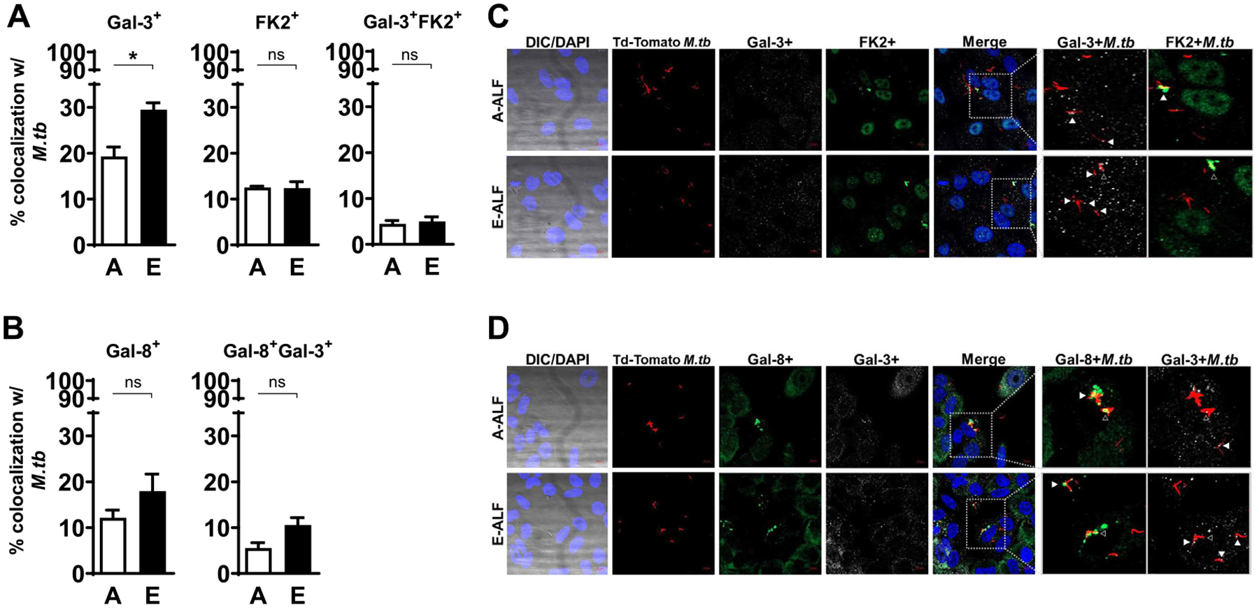
Fig. 6.

E-ALF exposure increases M.tb endosomal membrane damage within ATs. ATs were infected with either A-ALF or E-ALF-exposed Td-Tomato M.tb for 2 hours at MOI of 100:1 followed by 1 hour of gentamicin to kill extracellular M.tb. Monolayers were stained with different intracellular markers at 72 hpi. (A-B) Co-localization events indicative of compartment fusion of Mtb-Galectin-3+, Mtb-Galectin-8+, and M.tb-FK2+ (accumulation of ubiquitinated proteins), representing damaged endosomal membranes; n = 3–4 (mean ± SEM) in duplicate, using pooling of three different A-ALFs donors or four different E-ALFs. (C-D) Representative confocal images of ATs infected with A-ALF-exposed and E-ALF-exposed M.tb stained with intracellular markers Gal-3 and FK2 (C) or Gal-8 and Gal-3 (D) at 72 hours post-infection. Events were enumerated by counting 100–300 independent events (bacteria). The region indicated by the gray dashed line is shown expanded on the right; and co-localization events are indicated by white arrowheads. Open arrowheads indicate double co-localization events (Gal-3+FK2+ or Gal-8+Gal-3+). <scale bars represent 10 μm>. Student’s unpaired t test analysis of Adult versus Elderly, *p < 0.05. The “n” values represent the number of biological replicas using pooling of ALF samples from different adult or elderly human donors. A = adult ALF-exposed M.tb; ALF = alveolar lining fluid; ATs = alveolar epithelial type cells; DAPI = 4’,6-diamidino-2-phenylindole (ATs nuclear DNA); DIC = differential interference contrast; E = elderly ALF-exposed M.tb; FK2 = ubiquitinylated proteins (clone FK2); Gal = Galectin; hpi = hours post-infection; MOI = multiplicity of infection; M.tb = Mycobacterium tuberculosis; ns = no significant differences; SEM = standard error of the mean.
