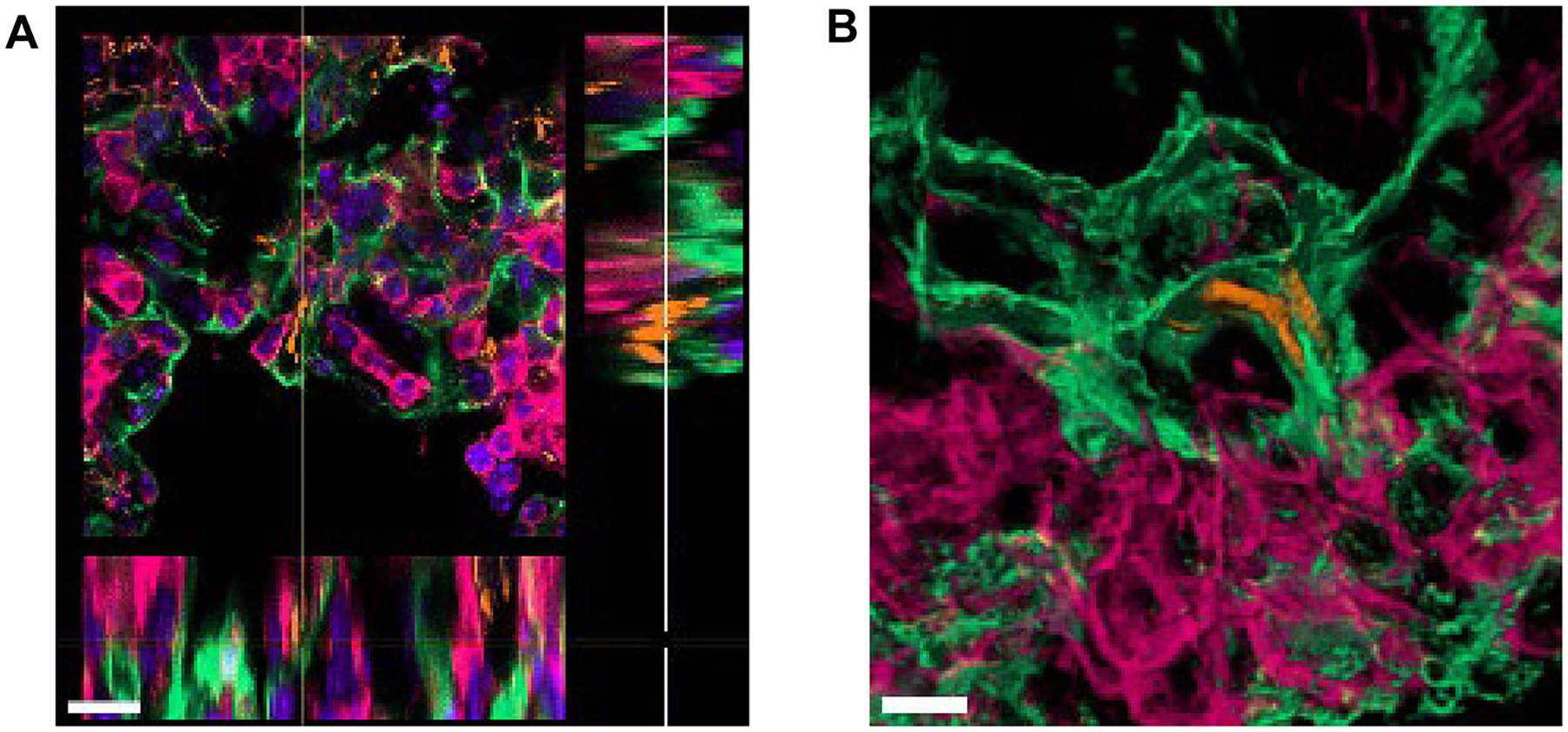
Fig. 9.

M.tb-infected ATs in early infection in the mouse model. (A) Extended ortho-section and (B) 3D view of M.tb bacilli intracellular in AT cells in the lungs of C57BL/6 mice infected with M.tb Erdman constitutively expressing Td-Tomato (orange) at 15 days post-infection. Podoplanin, PDPN (positive type I ATs, green); CD45 (leukocyte marker, pink), and DAPI (DNA/ nuclei marker, blue in panel A only). <scalebars (A) 15 μm, (B) 5 μm>. 3D = three-dimensional; ATs = alveolar epithelial type cells; DAPI = 4’,6-diamidino-2-phenylindole; DNA = deoxyribonucleic acid; M.tb = Mycobacterium tuberculosis; PDPN = podoplanin.
