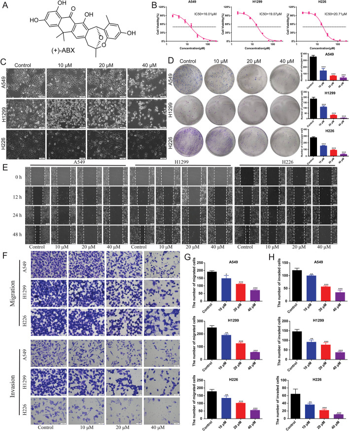
Fig. 1.
Inhibition of proliferation, migration, and invasion of NSCLC cell lines by (+)-ABX. A Chemical structure of (+)-ABX. B Determination of IC50 values using CCK-8 assay. C Morphological changes in cells treated with various concentrations of (+)-ABX for 24 h. D Colony formation of NSCLC cells after treatment with different concentrations of (+)-ABX. Colony-forming ability was quantified by counting colonies per well. E Measurement of wound healing in NSCLC cells after (+)-ABX treatment. F Assessment of transwell migration and invasion of NSCLC cells after (+)-ABX treatment. G Quantitative analysis of wound healing, transwell migration, and invasion assays. The bar graphs represent mean ± SD of at least three independent experiments; *P < 0.05, **P < 0.01, and ***P < 0.001 compared with the control group

