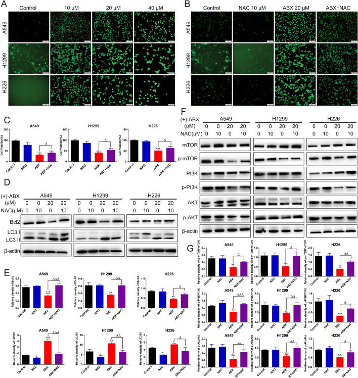
Fig. 7.
Enhanced ROS levels in NSCLC cells induced by (+)-ABX treatment. A Detection of ROS levels in cells treated with (+)-ABX using the DCFH-DA method. B Assessment of changes in ROS levels in cells treated with (+)-ABX after NAC intervention using the DCFH-DA method. C Impact of NAC intervention on cell viability after treatment with (+)-ABX measured by the CCK-8 assay. D Expression changes of apoptosis and autophagy-related proteins in cells treated with (+)-ABX after NAC intervention using western blotting. E Quantification of data from (D). F Expression changes of PI3K/AKT/mTOR pathway-related proteins in cells treated with (+)-ABX after NAC intervention using western blotting. G Quantification of data from (F). Data are presented as means ± SD. Statistical significance: * or △P < 0.05, ** or △△P < 0.01, and *** or △△△P < 0.001 compared with the control. Data were obtained from at least three independent experiments

