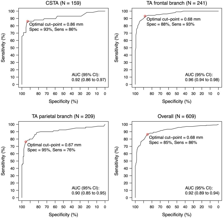
Figure 1.
ROC-curve for intima-media thickness for each temporal artery segment and overall. Intima-media thickness values are shown for the compressed lumen technique (combining both walls). The circle indicates the statistically optimal cut-off. Eleven Patients with giant cell arteritis had a normal T1-BB-MRI for all segments and were not included in this analysis. N indicates the number of segments (a maximum of two per patient, left and right side). AUC, area under the curve; CI, confidence interval; CSTA, common superficial temporal artery; IMT, intima-media thickness; ROC, receiver operating characteristic; TA, temporal artery.

