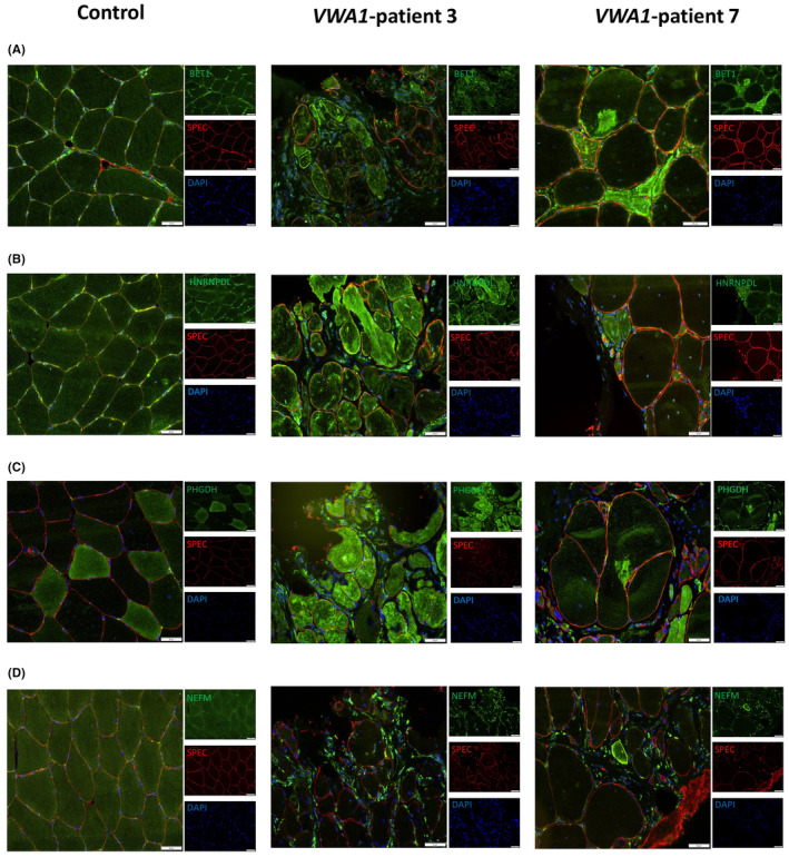
FIGURE 2.

Immunofluorescence findings in VWA1‐patient derived muscle biopsies. (A) Immunofluorescence studies of BET1 (green) showed a sarcoplasmic increase accompanied by the presence of focal dot‐like structures in patient‐derived muscle cells compared to muscle cells derived from a control case. Spectin (SPEC) staining (red) visualizes the sarcolemma. (B) Immunostaining of HNRNPDL also revealed a sarcoplasmic increase accompanied by the presence of dot‐like immunoreactive structures in patient‐derived muscle cells compared to control cells. Spectin (SPEC) staining (red) visualizes the sarcolemma. (C) VWA1‐mutant muscle cells show a generalized sarcoplasmic increase with the presence of focal accumulations of PHGDH (green) in comparison to non‐mutant muscle cells. Spectin (SPEC) staining (red) visualizes the sarcolemma. (D) Immunofluorescence‐based studies of NEFM revealed a sarcoplasmic increase only in few myofibres in addition to a considerable increase in extra muscular cells localized within the extracellular matrix in VWA1‐mutant muscle compared to controls. One representative control biopsy is shown for the different staining studies. Spectrin (SPEC) staining (red) visualizes the sarcolemma. DAPI staining visualizes nuclei. Scale bar 50 μm.
