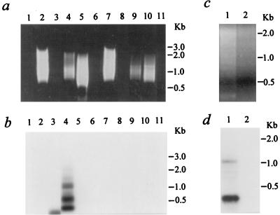
FIG. 3.
Alu-PCR analysis on 293 cell samples. (a and b) Cells were either untreated or transduced with a crude preparation of CMVβ-gal. (a) Aliquots of 25 μl of PCR products were loaded in a 1% agarose gel containing ethidium bromide. Lane 1, control sample without a genomic DNA template; lanes 2 to 6, 100 ng of genomic DNA isolated 8 days after transduction of the CMVβ-gal vector into 293 cells; lanes 7 to 11, 100 ng DNA from control 293 cells; lanes 2 and 7, 10 pmol of Alu5 primer alone for the first 10 cycles; lanes 3 and 8, 10 pmol of C1 primer alone; lanes 1, 4, and 9, Alu5-C1 at 10 pmol: 10 pmol; lanes 5 and 10, Alu5-C1 at 100:10; lanes 6 and 11, Alu5-C1 at 10:100. (b) The gel shown in panel a subjected to Southern blot analysis. PCR-amplified products on a nylon membrane were hybridized with a 32P-end-labeled specific CMV oligonucleotide probe (C3). (c and d) Alu-PCR amplification from 100 ng of genomic DNA isolated 8 days after transduction with CsCl gradient-purified CMVβ-gal. (c) Aliquots of 25 μl of PCR products loaded in an agarose gel. Lane 1, Alu5-C1 at 10 pmol: 10 pmol; lane 2, C1 primer alone. (d) The gel shown in panel c subjected to Southern blot analysis.

