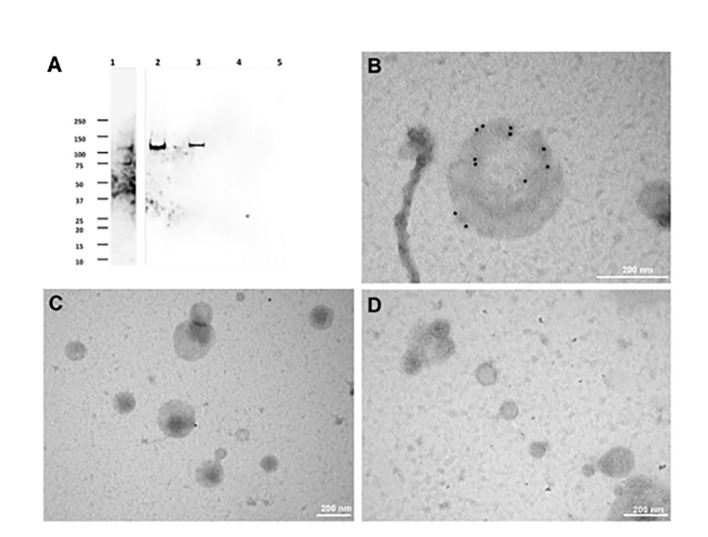
Figure 4.
Immunolocalization of CpRom1 by Western blotting on microvesicle extracts and immunoelectron microscopy of the corresponding extracellular vesicles. (A) Immunoblotting with anti-CpRom1 mouse serum: 1, unexcysted oocysts lysate; 2, excysted soporozoites; 3, LEVs extract; 4, SEVs extract; 5, TCA-precipitated supernatant of SEVs. (B) Negative staining immunoelectron microscopy of LEVs labelled with anti-CpRom1 rabbit serum. (C) Negative staining immunoelectron microscopy of LEVs labelled with pre-immune rabbit serum. (D) Negative staining immunoelectron microscopy of SEVs labelled with anti-CpRom1 rabbit serum.

