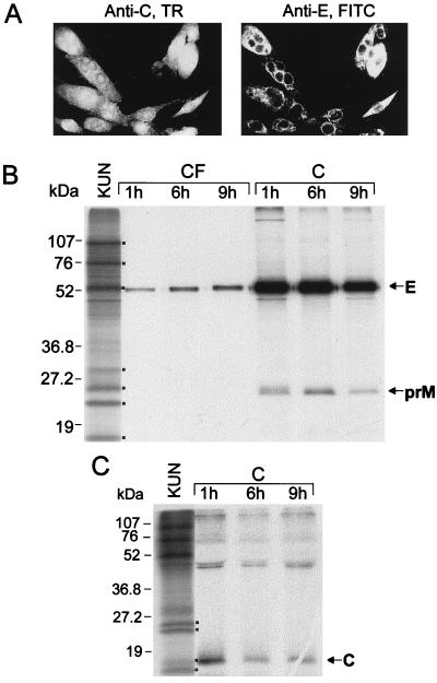
FIG. 4.
Expression of all three KUN structural proteins by the recombinant SFV-prME-C107 replicon. (A) Dual IF analysis of the same field of BHK21 cells fixed in acetone at 18 h after transfection with SFV-prME RNA by using KUN anti-C and anti-E antibodies with Texas red (TR)- and fluorescein isothiocyanate (FITC)-conjugated secondary antibodies, respectively. (B and C) Cells at 18 h after transfection with SFV-prME-C107 RNA were pulsed with [35S]methionine-cysteine for 1 h; subsequently, 300 μl (from a total of 600 μl) of cell lysates (lanes C) and 1 ml (from a total of 2 ml) of culture fluids (lanes CF) collected at different chase intervals (1, 6, and 9 h) were immunoprecipitated either with KUN anti-E antibodies (B) or with KUN anti-C antibodies (C) as described in Materials and Methods. Ten microliters (from a total of 30 μl) of immunoprecipitated samples was electrophoresed in SDS–12.5% (B) and –15% (C) polyacrylamide gels. Dots in panel B indicate KUN proteins in the labeled KUN cell lysates as in Fig. 2B. Dots in panel C represent KUN proteins prM, NS2A, C, and NS4A/NS2B (from top to bottom) in the radiolabeled KUN-infected cell lysate. Numbers represent sizes of the low-range prestained Bio-Rad protein standards.

