Figure 1.
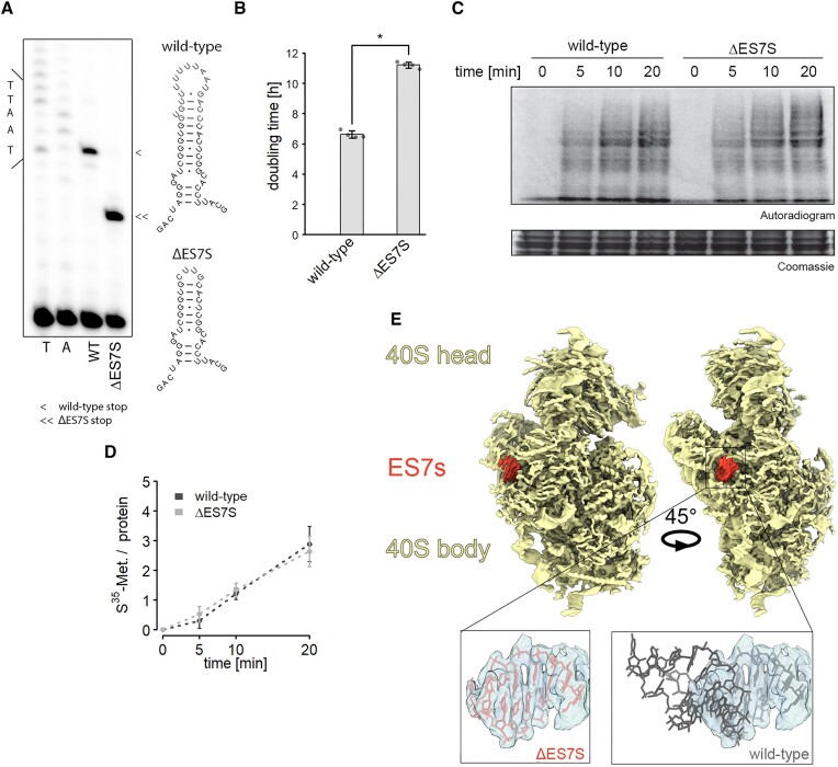
Ribosomal activity is unaltered in slow-growing ΔES7S cells. (A) Generation of pure ribosome populations was verified by poisoned primer extension assays. Reverse transcription products stop at different positions due to the successful removal of ES7S. The secondary structures display full-length ES7S on top and the truncated ΔES7S at the bottom. Arrowheads indicate stops due to the poisoned nucleotide. (B) Growth curves of wild-type and ΔES7S cells were measured in a Tecan plate reader and rates were calculated from the exponential growth phase. Stars indicate significant differences in growth rates (n = 4, P ≤ 0.05, Wilcoxon Rank Sum test). (C) In vitro translation was performed for the indicated times with lysates obtained from wild-type and ΔES7S cells. Reactions were electrophoresed and radiograms were imaged. (D) The radioactive signal was quantified and normalized to the protein loaded (n = 4, error bars indicate the standard deviation). (E) The structure of the small ribosomal subunit lacking ES7S was solved using cryo-EM. The rudimentary helix 26 is shown in red and the electron density for the helix is indicated in the zoom and compared to the wild-type structure.

