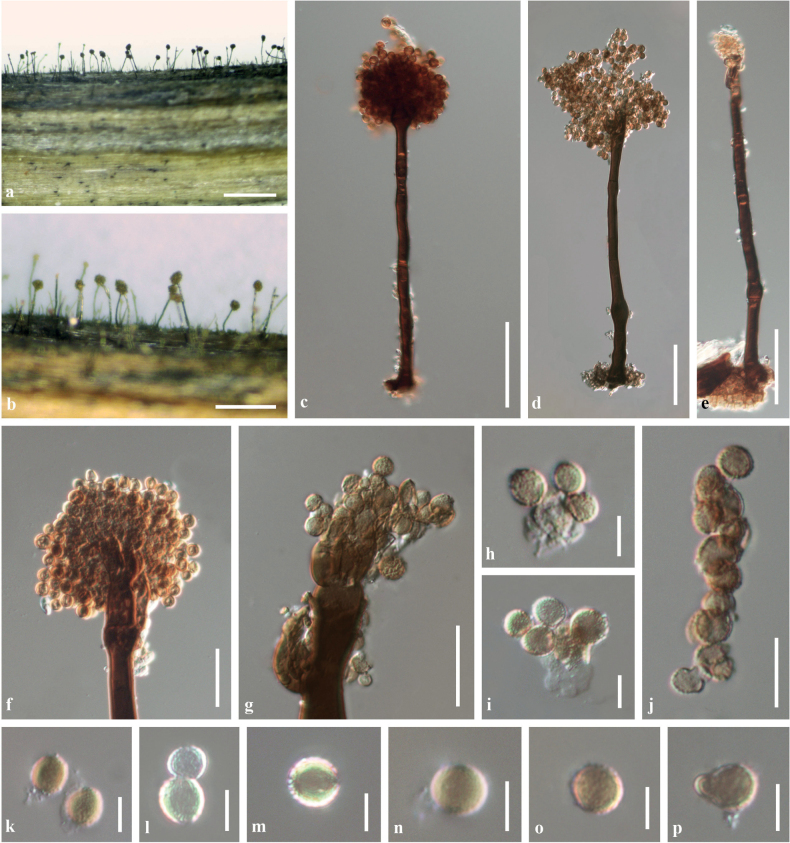
Figure 6.
Periconiakunmingensis (KUN-HKAS102239, holotype) A, B the appearance of fungal colonies on host substrate C–E conidiophores F, G closed-up conidiophores with spherical heads H, I conidiogenous cells bearing conidia J conidia catenate in acropetal short chain K–P conidia. Scale bars: 500 µm (A, B); 50 µm (C–E); 20 µm (F, G); 10 µm (J); 5 µm (H, I, K–P).

