
Figure 6.
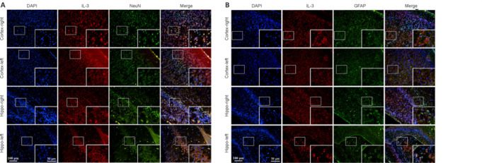
Double immunofluorescence staining of IL-3 and NeuN (A) or GFAP (B) in the right and left cortex and hippocampus.
(A) Double immunofluorescence staining of IL-3 and NeuN showed that the expression of IL-3 in neurons was obviously higher in the right cortex and hippocampus compared with that of the left cortex and hippocampus. The red (Dylight 594) indicates IL-3-positive cells, green (Dylight 488) indicates NeuN-positive cells (neurons), and blue represents DAPI (nuclei). Scale bars: 100 μm, 50 μm (enlarged image). (B) Double immunofluorescence staining of IL-3 and GFAP showed that the expression of IL-3 in astrocytes was obviously higher in the right cortex and hippocampus compared with that of the left cortex and hippocampus. Scale bars: 100 μm, 50 μm (enlarged image). The red (Dylight 594) indicates IL-3-positive cells, green (Dylight 488) indicates GFAP-positive cells (astrocytes), and blue represents DAPI (nuclei). DAPI: 4′,6-Diamidino-2-phenylindole; GFAP: glial fibrillary acidic protein; IL-3: interleukin-3; NeuN: neuron-specific nuclear protein.