
Figure 2.
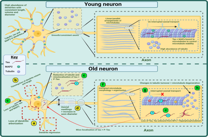
Graphical representation of age-related changes occurring to the morphology of the somatodendritic and axonal neuronal compartments and the cytoskeleton.
Evidence shows an age-related loss of and regression to dendrites (a) alongside a thinning and reduction in dendritic volume (b). Within the dendrites there is a loss of tubulin, tau, and MAP2 whilst tubulin is reduced in the soma and tau + P-Tau is mislocalized to the soma (c). In the axon, there can be increases in axonal diameter and the appearance of swellings (d). Normal tau is reduced and phosphorylated tau is increased whilst both also appear to mislocalize to the cell soma (e). EM studies show a disruption in the organization and morphology of microtubules (f) whilst axonal transport is interrupted (g) and GTPase derived tubulin turnover altered (h). Green circles represent MAP2 proteins, Red symbols represent tau proteins, purple circles represent tubulin, and blue (P) symbols represent phosphorylation. Created with BioRender.com. EM: Electron-microscopy; MAP: microtubule-associated protein; MTs: microtubules; p-Tau: phosphorylated tau.