
Figure 3.
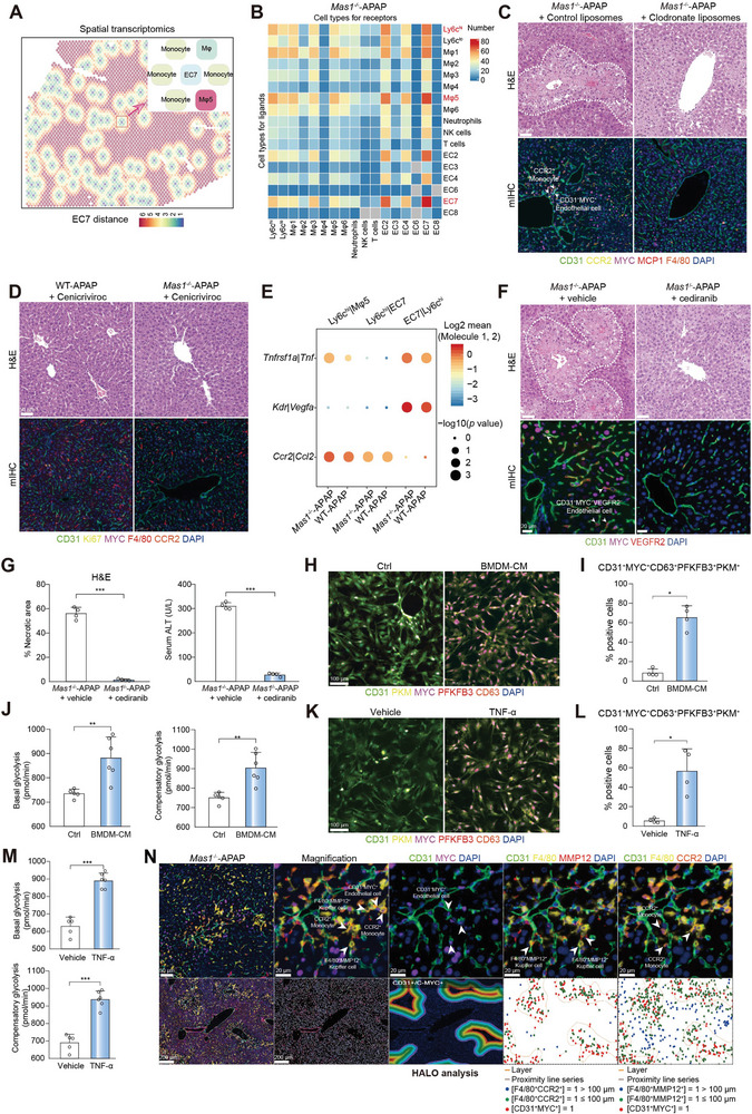
Monocyte‐derived TNF‐α acts on MYC+CD63+ EC through TNFR1. A) Spatial feature plots showing the cell types in EC7 expansion units for ST. B) CellPhoneDB analysis showing the cell–cell interactions for scRNA‐seq. C) Mas1 −/− mice were pre‐administrated with clodronate liposomes or control liposomes for 24 h before APAP challenge (n = 4 per group). Representative stainings of H&E and mIHC are shown. Scale bar: 50 µm. D) WT and Mas1 −/− mice were pre‐administrated with cenicriviroc for 2 h before APAP challenge (n = 4 per group). Representative stainings of H&E and mIHC are shown. Scale bar: 50 µm. E) CellPhoneDB analysis showing the major interaction pairs between Ly6chi monocyte‐Mψ5 or Ly6chi monocyte‐EC7 for scRNA‐seq. In F‐G, Mas1 −/− mice were pre‐administrated with cediranib or control for 2 h before APAP challenge (n = 4 per group). F) Representative staining of H&E and mIHC are shown. Scale bar: 50 µm. G) Quantification of necrotic area for H&E as shown in F (n = 4 mice per group; two‐sided Student's t‐test; p = 1.55 × 10⁻⁴) and serum ALT (n = 4 mice per group; two‐sided Student's t‐test; p = 2.64 × 10⁻⁸). H) Multiplex fluorescence of CD31+MYC+CD63+ LSECs and key glycolytic enzymes (PKM and PFKFB3). Scale bar: 100 µm. The mouse primary LSECs were treated with or without BMDM‐CM for 24 h. I) Quantification of CD31+MYC+CD63+PFKFB3+PKM+ LSECs for multiplex fluorescence as shown in H (n = 4 samples per group; two‐sided Mann‐Whitney U test, p = 2.86 × 10⁻²). J) Glycolytic rate assay showing glycolytic function of LSECs treated with or without BMDM‐CM for 24 h. (n = 5 or 6 samples per group; two‐sided Student's t‐test, p = 7.50 × 10⁻³ and 3.77 × 10⁻³ from left to right). K) Multiplex fluorescence of CD31+MYC+CD63+ LSECs and key glycolytic enzymes (PKM and PFKFB3). Scale bar: 100 µm. The mouse primary LSECs were treated with or without TNF‐α (20 ng ml−1) for 24 h. L) Quantification of CD31+MYC+CD63+PFKFB3+PKM+ LSECs for multiplex fluorescence as shown in K (n = 4 samples per group; two‐sided Student's t‐test, p = 2.00 × 10⁻²). M) Glycolytic rate assay showing glycolytic function of LSECs treated with or without TNF‐α (20 ng ml−1) for 24 h. (n = 5 or 6 samples per group; two‐sided Student's t‐test, p = 8.00 × 10⁻⁶ and 1.30 × 10⁻⁵ from top to bottom). N) mIHC of CD31+MYC+ ECs, F4/80+MMP12+ Mψ and CCR2+ monocytes are shown. Scale bar: 200 µm, 50 µm and 20 µm. Spatial localizations of CD31+MYC+ ECs, CCR2+ monocytes, and F4/80+MMP12+ Mψ within injured region are shown. In all graphs data are presented as mean ± SD, *p < 0.05; **p < 0.01; ***p < 0.001.