Figure 1.
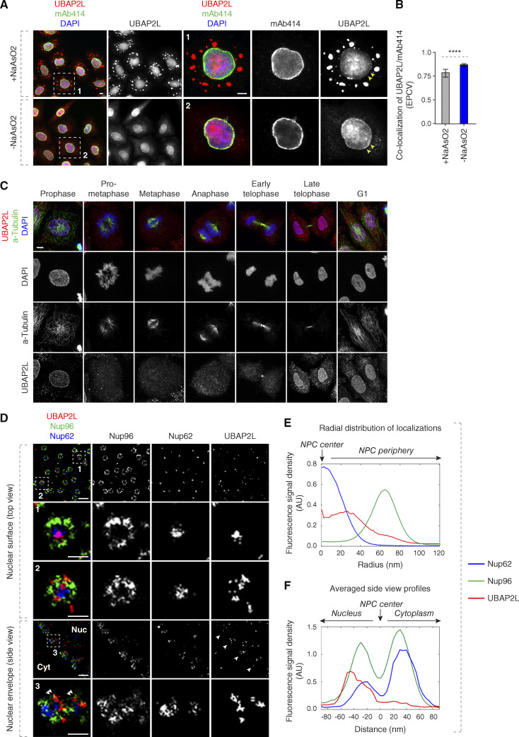
UBAP2L localizes to the NE and NPCs. (A and B) Representative images of the localization of UBAP2L and Nups in HeLa cells with/without NaAsO2 treatment shown by immunofluorescence microscopy with UBAP2L and mAb414 antibodies. Nuclei were stained with DAPI. The arrowheads indicate the NE localization of endogenous UBAP2L. The magnified framed regions are shown in the corresponding numbered panels. Scale bars, 5 μm (A). The colocalization (EPCV, events per cell view) of UBAP2L and mAb414 in A was measured by CellProfiler (mean ± SD, ****P < 0.0001, unpaired two-tailed t test; 175 cells for NaAsO2 treatment and 110 cells without NaAsO2 treatment were counted) (B). (C) Representative immunofluorescence images depicting the localization of UBAP2L in HeLa cells after chemical pre-extraction of the cytoplasm using 0.01% of Triton X-100 for 90 s in indicated cell cycle stages and visualized by UBAP2L antibody. Nuclei and chromosomes were stained with DAPI. Scale bar, 5 μm. (D–F) Representative super-resolution immunofluorescence images of Nup96-GFP KI U2OS cells acquired using multicolor SMLM with a dichroic image splitter (splitSMLM) show NPCs on the nuclear surface (top view) and in the cross-section of the NE (side view). Nup96 signal labels the cytoplasmic and nuclear ring of the NPC and the localization of the central channel NPC component is analyzed by Nup62 antibody. The nuclear (Nuc) and cytoplasmic (Cyt) sides of the NE are indicated in the side view. The magnified framed regions are shown in the corresponding numbered panels. Note that UBAP2L can localize to both structures within the NPCs (framed regions 1 and 2 in the top view) and is found preferentially at the nuclear ring labeled with Nup96 (double arrowheads in framed region 3 in the side view). Scale bars, 300 and 100 nm, respectively (D). Radial distribution of localizations of Nup62, Nup96, and UBAP2L in D was obtained by averaging 1932 NPC particles (E). Averaged “side view” profiles of Nup62, Nup96, and UBAP2L in D were obtained by alignment of 83 individual NPCs (F). Orientation bars point to the NPC center (central channel middle point) as well as the cytoplasmic and nuclear sides (E and F).

