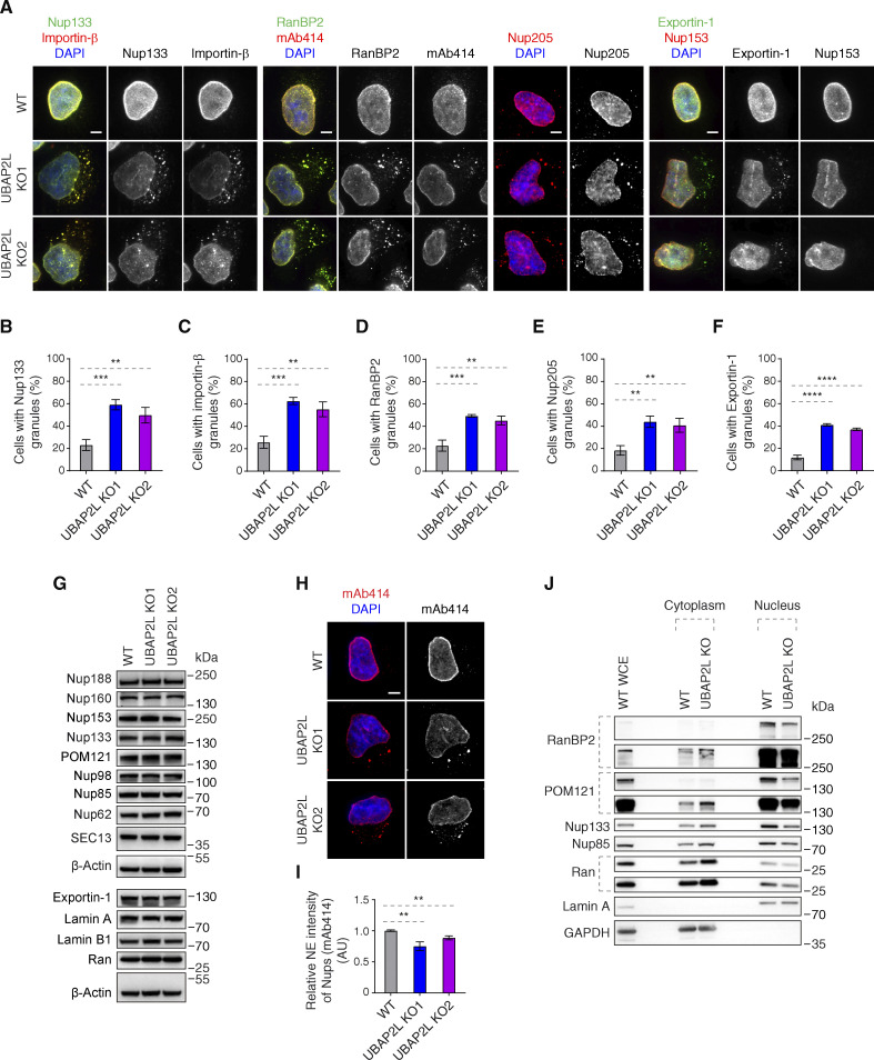
Figure 3.
UBAP2L regulates Nups localization. (A–F) Representative immunofluorescence images depicting the localization of Nups and NPC-associated factors in WT and UBAP2L KO HeLa cells synchronized in interphase by DTBR at 12 h. Nuclei were stained with DAPI. Scale bars, 5 μm (A). The percentage of cells with the cytoplasmic granules containing Nup133 (B), Importin-β (C), RanBP2 (D), Nup205 (E), and Exportin-1 (F) in A were quantified. At least 100 cells per condition were analyzed (mean ± SD, **P < 0.01, ***P < 0.001, ****P < 0.0001, unpaired two-tailed t test, n = 3 independent experiments). (G) The protein levels of Nups and NPC-associated factors in WT and UBAP2L KO HeLa cells synchronized in interphase by DTBR at 12 h were analyzed by western blot. (H and I) Representative immunofluorescence images of FG-Nups (mAb414) at the NE in WT and UBAP2L KO HeLa cells in interphase cells synchronized by DTBR at 12 h. Nuclei were stained with DAPI. Scale bar, 5 μm (H). The NE intensity of Nups (mAb414) in H was quantified (I). At least 150 cells per condition were analyzed (mean ± SD, **P < 0.01, unpaired two-tailed t test, n = 3 independent experiments). (J) The nuclear and cytoplasmic protein levels of Nups and NPC transport-associated factors in WT and UBAP2L KO HeLa cells synchronized in the G1/S transition phase by thymidine 18 h were analyzed by western blot. WCE indicates whole cell extract. Source data are available for this figure: SourceData F3.

