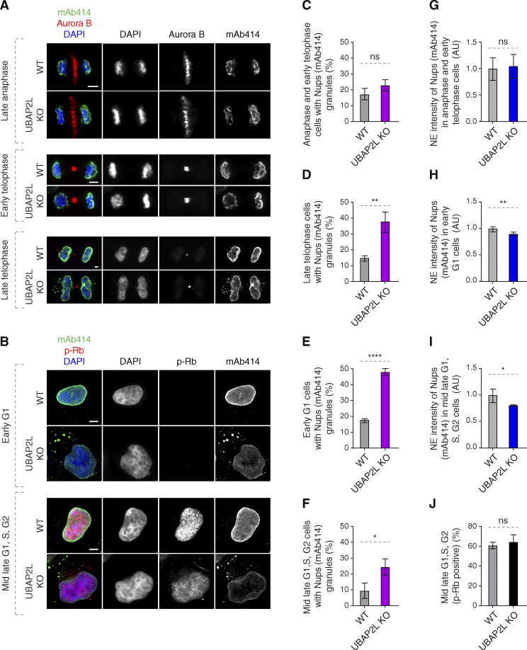
Figure 4.
UBAP2L regulates localization of Nups in interphase but not during mitotic exit. (A and B) Representative immunofluorescence images depicting the localization of Nups (mAb414) in WT and UBAP2L KO HeLa cells in different cell cycle stages. Mitotic cells were labeled by Aurora B (A) while p-Rb was used to distinguish between early G1 (p-Rb–negative cells) and mid-late G1, S, and G2 (p-Rb–positive cells) stages (B). Nuclei and chromosomes were stained with DAPI. Scale bars, 5 μm. (C–F) The percentage of cells with the cytoplasmic granules of Nups (mAb414) in anaphase and early telophase (C), late telophase (D), early G1 (E), and mid-late G1, S, G2 (F) in A and B were quantified. At least 150 cells per condition were analyzed (mean ± SD, ns: non-significant, *P < 0.05, **P < 0.01, ****P < 0.0001, unpaired two-tailed t test, n = 3 independent experiments). (G–I) The NE intensity of Nups (mAb414) in anaphase and early telophase cells (G), early G1 cells (H), and mid-late G1, S, G2 cells (I) in A and B were quantified. At least 100 cells per condition were analyzed (mean ± SD, ns: non-significant, *P < 0.05, **P < 0.01, unpaired two-tailed t test, n = 3 independent experiments). (J) The percentage of p-Rb–positive cells in B was quantified. At least 150 cells per condition were analyzed (mean ± SD, ns: non-significant, unpaired two-tailed t test, n = 3 independent experiments).

