Figure 6.
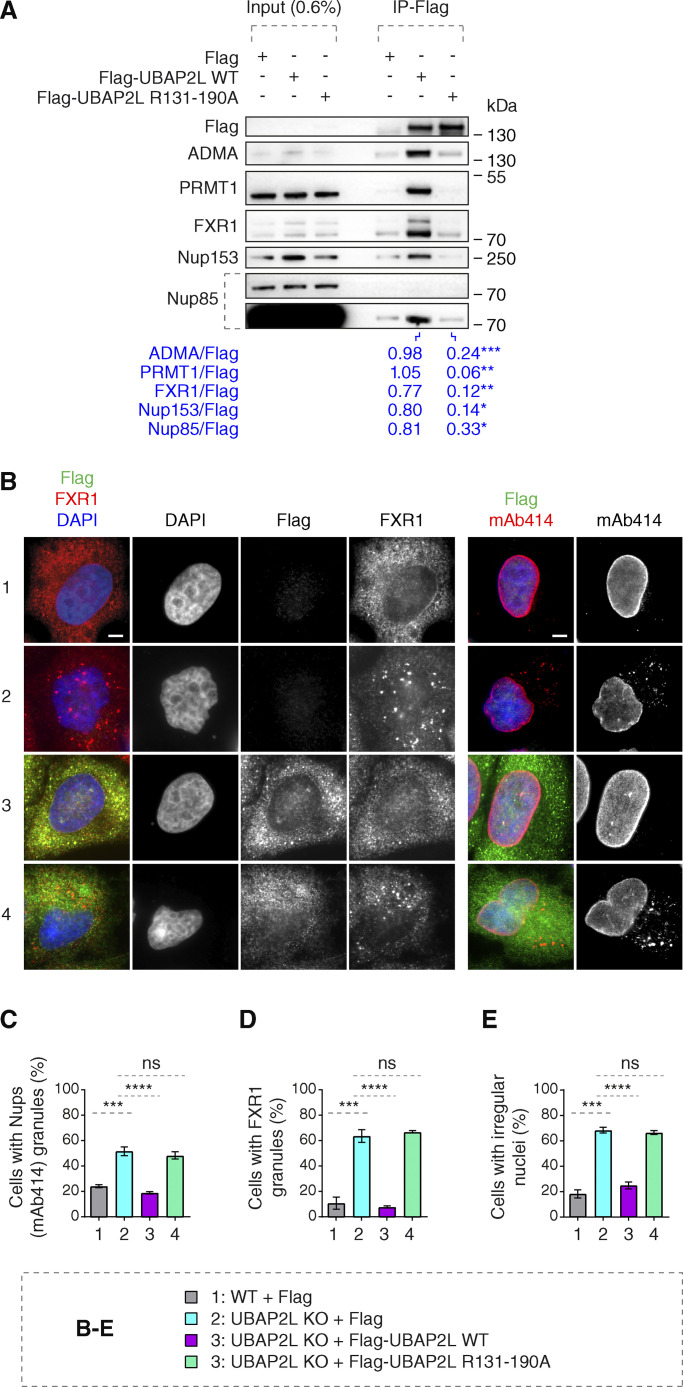
Arginines within the RGG domain of UBAP2L mediate the function of UBAP2L on Nups and FXRPs. (A) Lysates of interphase HeLa cells expressing Flag alone, Flag-UBAP2L WT, or mutated Flag-UBAP2L version where 19 arginines located in the RGG domain were replaced by alanines (R131–190A) for 27 h were immunoprecipitated using Flag beads (Flag-IP), analyzed by western blot, and signal intensities were quantified (shown a mean value, *P < 0.05, **P < 0.01, ***P < 0.001, unpaired two-tailed t test; n = 3 independent experiments). (B–E) Representative immunofluorescence images depicting nuclear shape and localization of FXR1 and Nups (mAb414) in WT and UBAP2L KO HeLa cells expressing Flag alone or Flag-UBAP2L (WT or R131–190A) for 60 h and synchronized in interphase by DTBR at 12 h. Nuclei were stained with DAPI. Scale bars, 5 μm (B). The percentage of cells with the cytoplasmic granules of Nups (mAb414) (C) and of FXR1 (D) and irregular nuclei (E) shown in B were quantified. At least 200 cells per condition were analyzed (mean ± SD, ns: not significant, ***P < 0.001, ****P < 0.0001, unpaired two-tailed t test, n = 3 independent experiments). Source data are available for this figure: SourceData F6.

