Figure 7.
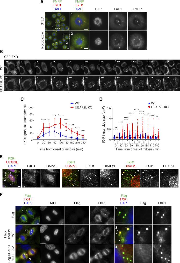
UBAP2L remodels FXR1-protein assemblies in the cytoplasm and drives localization of FXR1 to the NE. (A) Representative immunofluorescence images depicting the localization of FXR1 and FMRP in HeLa cells synchronized in prometaphase using STCL 16 h or nocodazole 16 h. Chromosomes were stained with DAPI. Scale bars, 5 μm. (B–D) WT and UBAP2L KO HeLa cells expressing GFP-FXR1 were synchronized by DTBR and analyzed by live video spinning disk confocal microscopy. The selected representative frames of the movies are depicted, and time is shown in minutes. Timepoint 0 indicates mitotic entry during prophase. Scale bar, 5 μm (B). GFP-FXR1 granules number (number/cell) shown in B at indicated times during mitotic progression were quantified (C). GFP-FXR1 granules sizes (granule ≥ 0.061 µm2) shown in B at indicated timepoints during mitotic progression were quantified (D). 16 WT and 11 UBAP2L KO HeLa cells were counted in C and D, respectively (mean ± SD, *P < 0.05, **P < 0.01, ****P < 0.0001, unpaired two-tailed t test ). (E) Representative immunofluorescence images depicting the cytoplasmic and NE localization of endogenous UBAP2L and FXR1 in interphase HeLa cells. Nuclei were stained with DAPI. The magnified framed regions are shown in the corresponding numbered panels. The arrowheads indicate co-localization of UBAP2L and FXR1 foci in the cytoplasm. Scale bar, 5 μm. (F) Representative immunofluorescence images depicting the localization of FXR1, Flag alone, and Flag-UBAP2L (WT or R131–190A) in late telophase in HeLa cells. Nuclei were stained with DAPI. The magnified framed regions are shown in the corresponding numbered panels. Note that Flag-UBAP2L WT (arrowheads) but not Flag alone and Flag-UBAP2L R131–190A is localized to FXR1 containing granules in proximity of NE. Scale bar, 5 μm.

