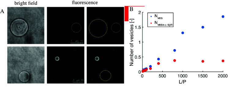
Figure 2.
Characterization of α-synuclein association with lipid membranes in terms of binding cooperativity based on experimental observation of protein distribution in excess of lipid membranes. (A) Nonrandom distribution of α-synuclein in a population of giant unilamellar vesicles (GUVs). Two sets of bright-field (left) and fluorescence (middle) images of nonfluorescently labeled GUVs and fluorescently labeled α-synuclein. The protein association with GUVs renders them visible in the fluorescence images. The protein-free GUVs invisible in the fluorescence images are marked with a yellow dotted circle in the rightmost panel. (B) Distribution of α-synuclein in a population of small unilamellar vesicles studied with fluorescence correlation spectroscopy (FCS). Blue dots represent the total number of vesicles in the sample, while red dots represent the number of vesicles having α-synuclein bound. The numbers were extracted from the amplitudes of the FCS autocorrelation functions. Reproduced from ref (43). Copyright 2021 American Chemical Society.

