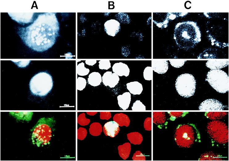
FIG. 4.
Subcellular localization of HCV core protein. RK13 cells (A) and HPB-Ma cells (B) were infected with both the LO-R6J20 and the LO-T7-1 RVVs. Cells were fixed with acetone and methanol at −20°C for 10 min 12 h after infection, counterstained with FITC-conjugated anti-mouse Ig (for HCV core protein) and propidium iodide (for nuclear DNA), and viewed with a confocal laser scanning microscope. (C) CHO cells stably transformed with an HCV core protein expression vector. Upper panels, HCV core protein; middle panels, nuclear DNA; lower panels, merged image of HCV core protein (green) and nucleus (red).

